Energy filtered PEEM
The Imaging Energy Filter (IEF) makes the FOCUS PEEM a powerful tool to measure work function maps (with lab sources) as well as element specific maps (with synchrotron, X-ray microspectroscopy) of conductive samples. For time/energy resolved measurements paired with a pulsed excitation source the FOCUS PEEM can be equipped with a Time-of-Flight (TOF) energy filter. All these options can be combined in one instrument for maximum versatility.
The PEEM with IEF and/or TOF combined with the optional angular imaging optics delivers laterally resolved k-space images (momentum microscopy).
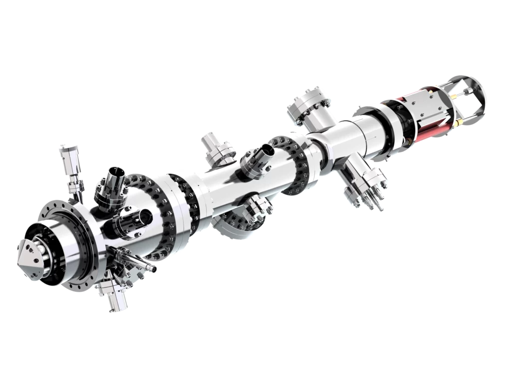
IEF PEEM & VUV lab source HIS 14 HD
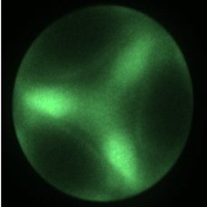
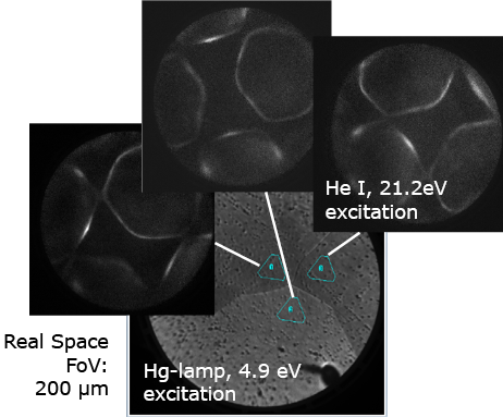
Imaging of the d-band region of Ag(111), He I excitation (HIS 14 HD, 21.2 eV), E-EF = 15.5 eV, dwell time 5 s, FoV = 20 μm
Right: µ-ARPES of polycrystalline silver. The navigation in the real space is accomplished by use of the iris micro aperture defining the region of interest before switching to k-space imaging.
Because of their different crystallographic orientations the single grains deliver different momentum patterns.