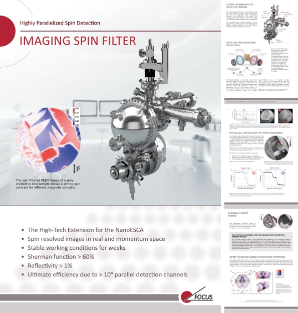
Module for NanoESCA III
The Imaging Spin Filter is available as module for the NanoESCA III analyzer. The monochromatic image delivered by the NanoESCA is a prerequisite for the defined kinetic electron energy needed for the spin-sensitive scattering process at the Au/Ir crystal.
Key features
- Stable scattering conditions for weeks
- Sherman function > 60%
- Reflectivity > 1%
- > 104 parallel detection channels
Further details
For an overview of our EFM Series please check out our in-depth brochure!
Spin Filter working principal
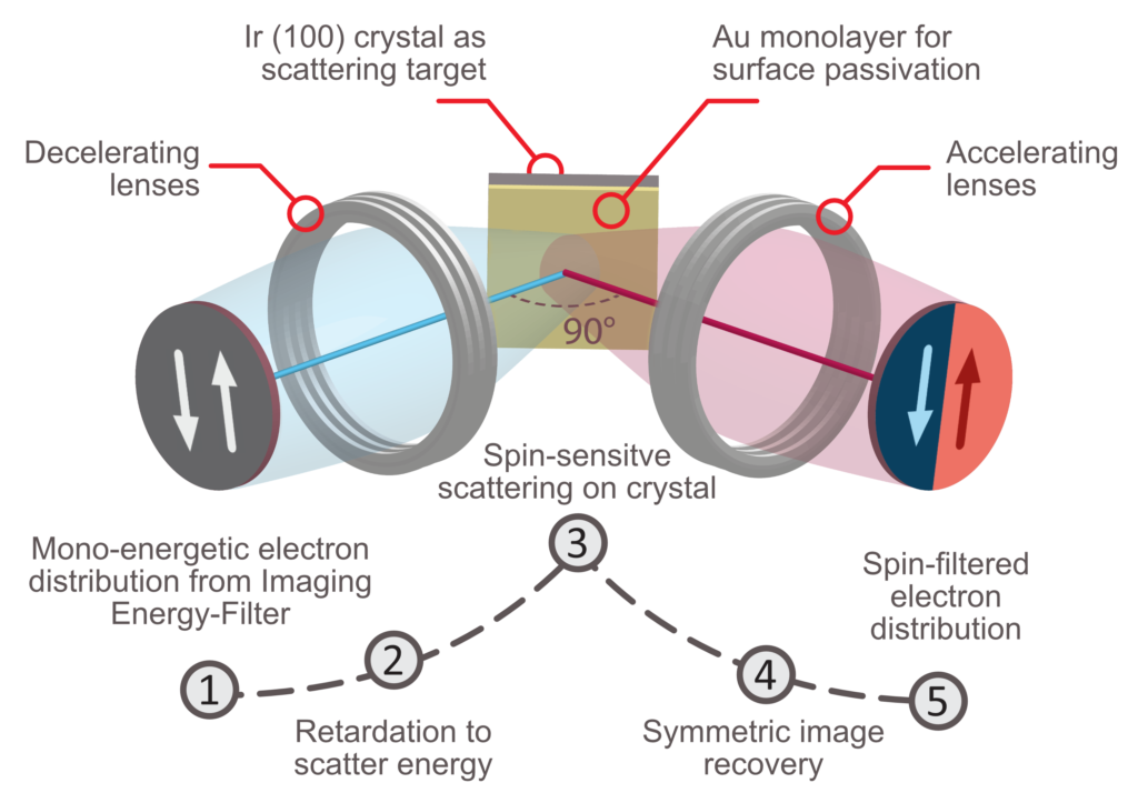
The monochromatic image delivered by the Imaging Energy-Filter of the NanoESCA is projected onto a Au/Ir crystal. For specific kinetic electron energies, one spin polarization is strongly preferred in the scattering process due to spin-orbit coupling. The reflected image is spin-filtered. The Au passivated surface of the Ir crystal keeps the scattering conditions stable for weeks.
protecting German Patents: DE102005045622B4, legal and technical rights secured by sublicensing from the Surface Concept GmbH
Pre-characterizations of the Au/Ir crystal were done with LEED and a Ferrum-Detector setup [1] to find optimal preparation conditions and scattering energies for a high single-point figure-of-merit (with Spin-sensitivity >60% and Reflectivity >1%) [2]. The I-V-curve in (a) was measured with the Ferrum setup and shows, at which scattering energy the highest spin-sensitivity can be reached. Images (b,c) are then acquired at these two energetically sharp working points with the NanoESCA and are used to calculate the spin-polarization of the complete field-of-view.
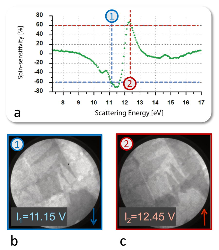
[1] Escher, et al., e-J. Surf. Sci. Nanotech. Vol. 9, 340-343 (2011)
[2] C.Tusche et al., Ultramicroscopy 159, 520–529 (2015)
Parallel detection of spin-channels
The already high single-point figure-of-merit (Sherman function > 60% and Reflectivity > 1%) is multiplied by the amount of parallel detection channels given in the imaging spin-filter to define an effective 2D figure-of-merit. It is 4 orders of magnitude larger than the figure-of-merit of single-point detectors.
The spin filtered PEEM image of a poly-crystalline iron sample shows a strong spin contrast for different magnetic domains.
- Field of View = 996 pixel along diameter
- Resolution of domain boundary = 7 pixel
- 142 resolvable image points (along diameter)
- π·(142/2)² = 15900 parallel detection channels
Measured with: NanoESCA, ELI-ALPS, Hg-discharge lamp, 100 meV instrumental energy resolution, Room temperature, 200 s exposure time, 66 µm lateral FoV
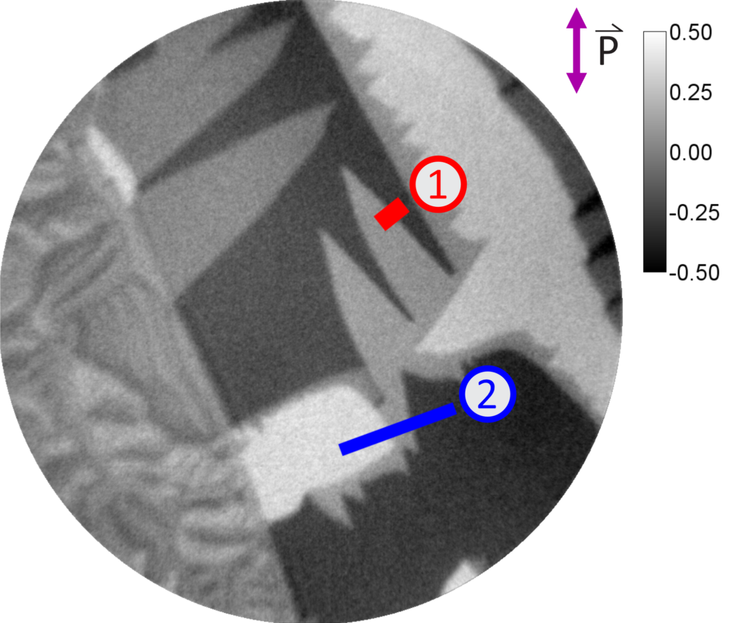
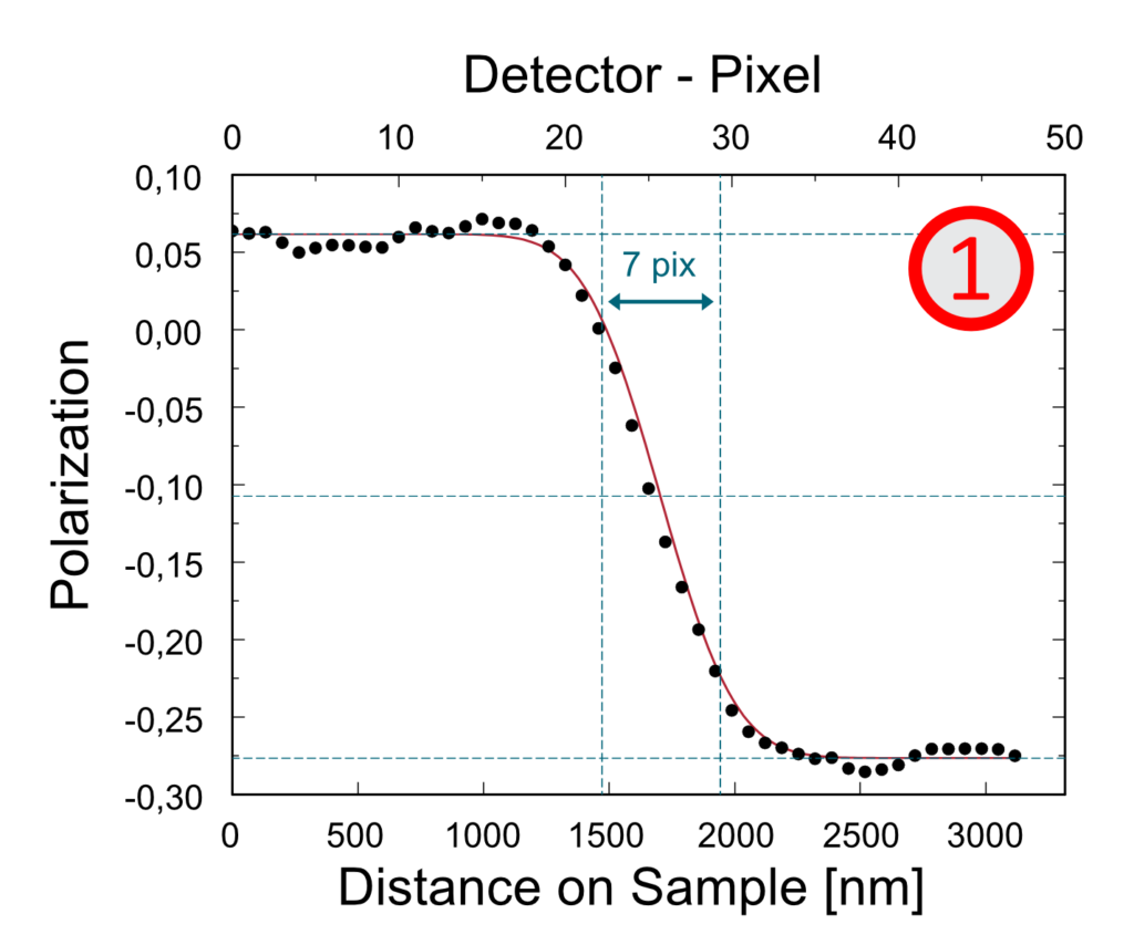

The line profiles show the spin polarization of different domains. The edge resolution reveals 142 resolvable image points along the diameter which corresponds to more than 104 parallel detection channels for the detector.
Spin Filtered band structure mapping
The Imaging Spin-Filter works in the momentum microscopy mode in the same way as in the real-space microscopy mode. The graphic shows the spin-resolved Fermi surface of W(110) (a) as well as full 3D spin-resolved momentum maps (momentum vs. binding energy) (b). Note, that a W (100) crystal was used as scattering target in this experiment.
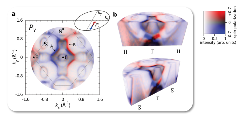
Measured with:
NanoESCA, Elettra (Trieste, Italy), Synchrotron radiation, hν = 50 eV, p-polarized, T = 130 K
Adapted from: Ying-Jiun Chen, Christian Tusche et al., Comm. Phys. (2021) 4:179 (CC-by 4.0 License)
Abstract: https://www.sciencedirect.com/science/article/pii/S0304399123001316