Time of Flight – Momentum and Real Space Imaging Spectroscopy
- Momentum and Real-Space Microscopy
- Time of Flight – Spectrometer with Energy Resolution < 30 meV
- Lateral Resolution < 35 nm
- Momentum-Space Resolution < 5 mÅ-1 for Band structure dynamics
- Dual Detector setup for fast and easy work flow (with High Pass Filter)
- Available with Normal Incidence Mirror
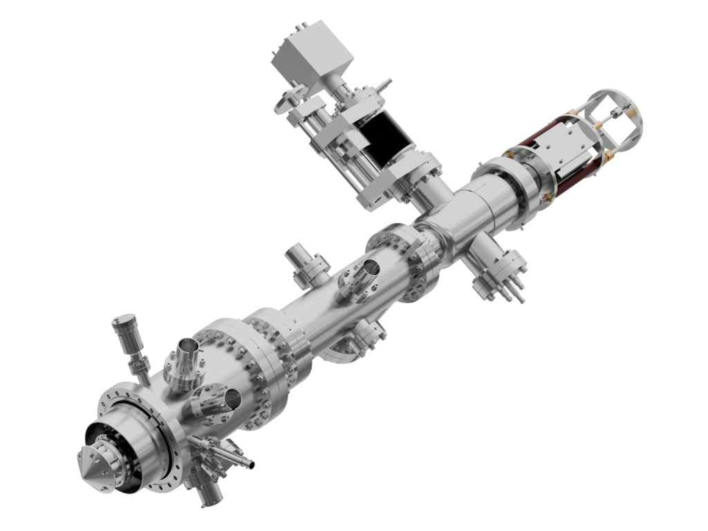
A TOF – PEEM with the new MARIS microscopy lens
Our Time-of-flight Photoemisson Electron Microsocope (TOF-PEEM) combines a microscopy lens with a special energy filter, in which the kinetic energy of photoelectrons is separated by their flight-time through a drift tube. The time-resolved and position sensitive delay-line detector (DLD) measures the arrival time of an electron, and references it to the light-pulse, which excited it.
When operated with pulsed light-sources, an energy-window of laterally or angle resolved photo electron distributions can be measured in parallel. That is often a more efficient alternative to hemispheres, which accomplish the energy separation by a small slit and a dispersive field.
FOCUS has a long history of improving this technology, starting with the first lateral-resolved PEEMs in 1995 and publishing the first scientific angle and energy-resolved PEEM results in 2002 (nowadays often referred to as Momentum Microscope). Our first TOF-PEEM with lateral and momentum resolution was build in 2007. The latest revision of our microscope lens was launched in 2022 in combination with a double-hemispherical energy-filter, called NanoESCA MARIS. Besides its high lateral resolution of 35 nm we could improve the momentum resolution by a factor of 3 to reach an unsurpassed momentum resolution of < 0.005 Å-1.
The same lens in combination with a time-of-flight energy filter complements our microscopy product line as TOF MARIS.
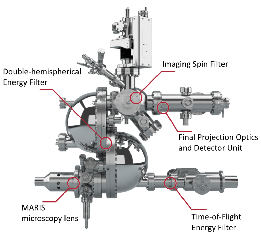
Instrumental Layout
The TOF MARIS utilizes two different detectors for an efficient work-flow. The Standard Imaging Unit (MCP + CCD Camera) is used for high count rates (> 30 Mcounts/s) and is perfectly suited to use continous lightsources with a high flux for the initial sample navigation in live view mode, e.g. to find interesting features on the sample under investigation.
Subsequently, the retractable delay-line-detector is moved into the optical axis and used for the time- / energy-resolved measurements with a pulsed light-source.
The microscope can be easily switched between real-space and momentum space projection with the projective lenses before the energy filter.
Parallel energy detection and energy – resolution
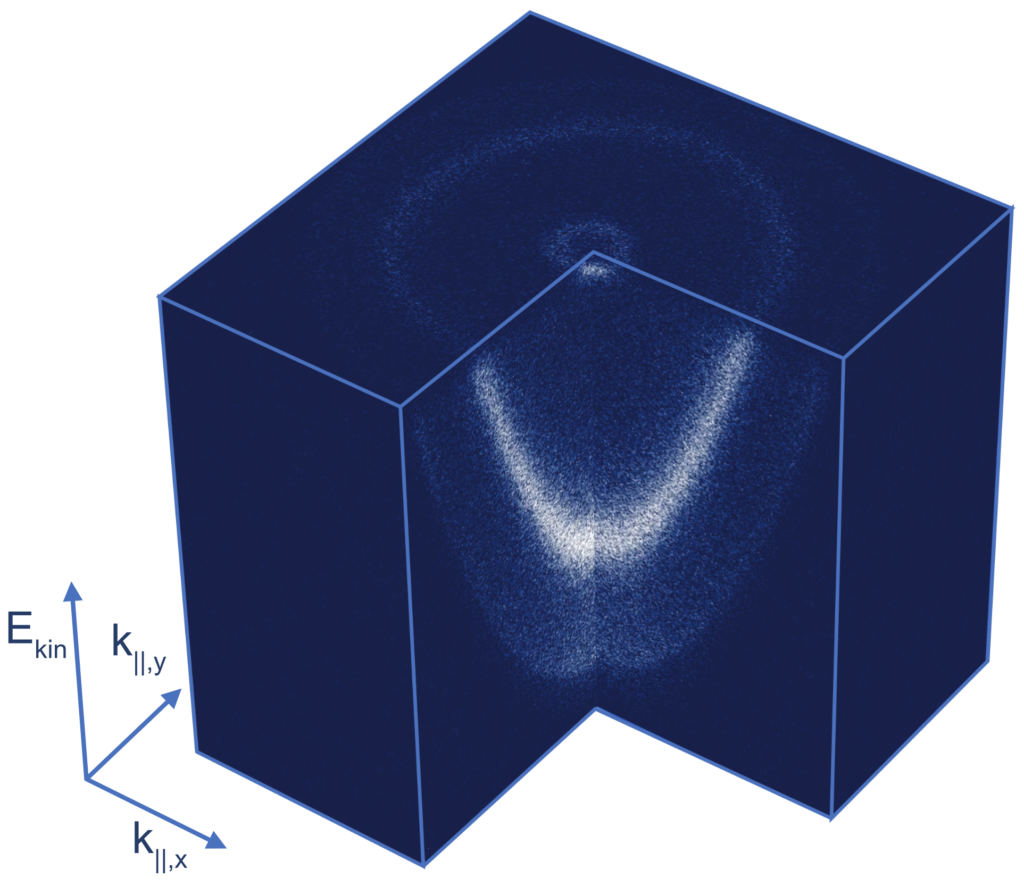
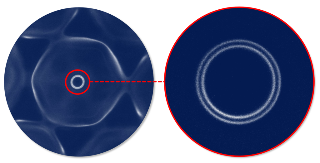
The benefit of a PEEM with time-of-flight energy filter is the parallel detection of the angular (or lateral) electron distribution and a large energy window. The data cube on the left was acquired in such a way. It shows the momentum vs energy measurement of a Ag (111) crystal, excited with a laser with a 3.1 eV photon energy.
With a 2 PPE process, electrons between the work function cut-off (E – EF = 4.4 eV) and the Fermi Level (E – EF=6.2 eV) can be acquired. The cube was measured at room temperature.
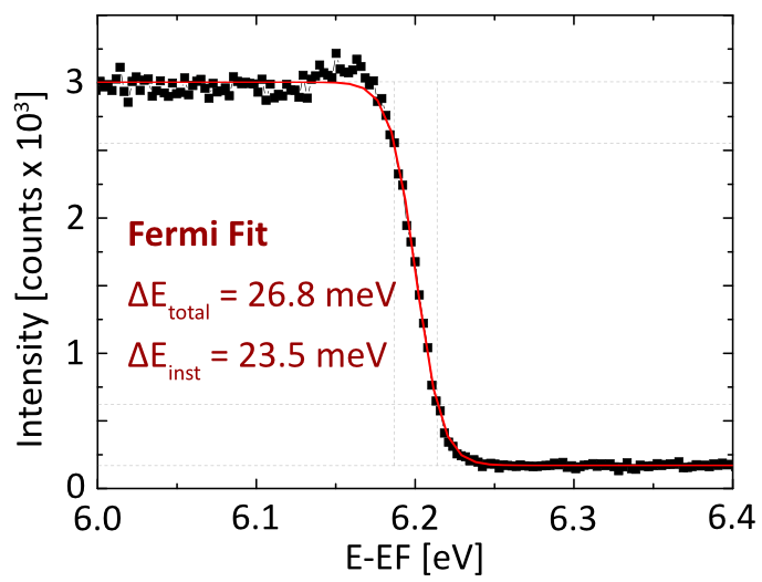
The Fermi Edge on the right was acquired with the same set-up, but a sample temperature of 45 K. It shows an instrumental energy resolution of ∆Einst = 23.5 meV.
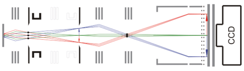
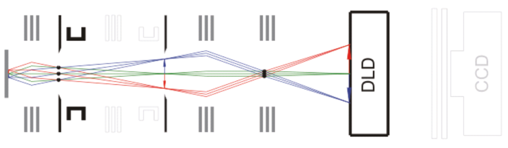
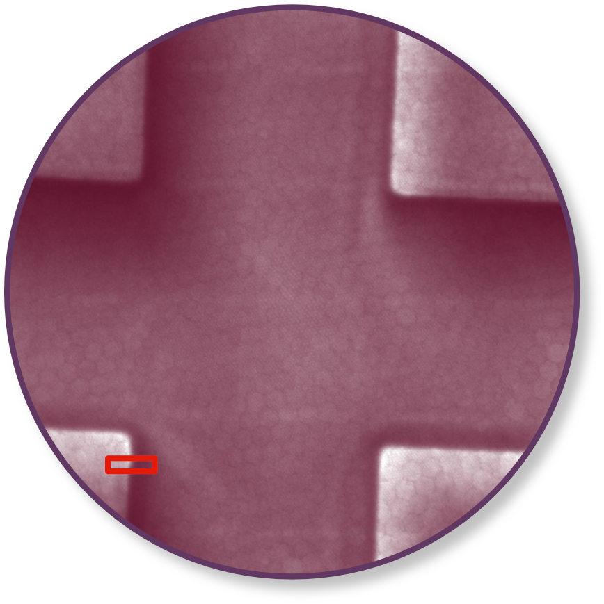
Lateral resolution with Standard Imaging Unit and DLD
Standard Imaging Unit
Delay Line Detector
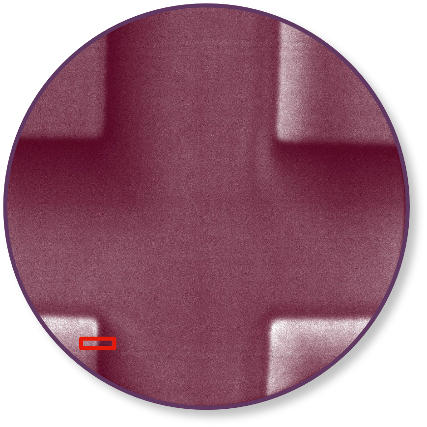
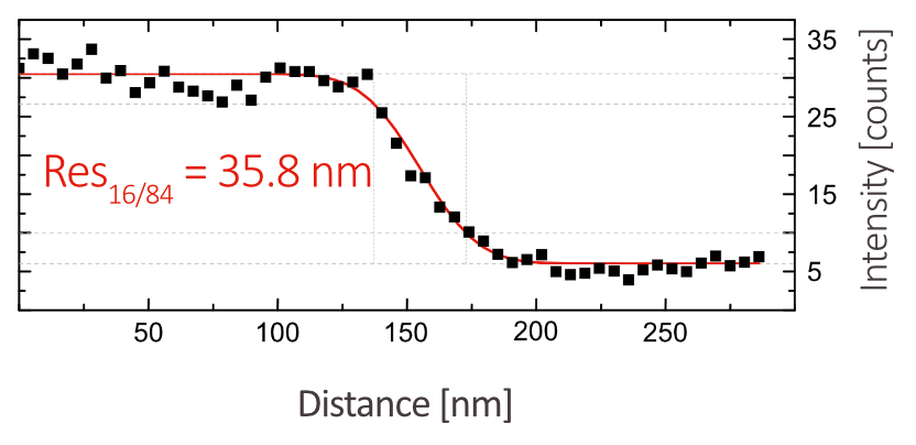
The two raw images were measured on the same region of a structured test sample (Si grid covered with Ag) with two different detectors. The image quality and resolutions are comparable. In both cases, a mercury arc lamp was used for photo electron excitation.
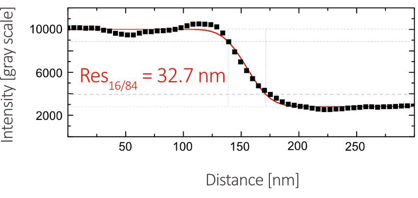
The line scans along the marked edges of the structures were fitted with a convolution of a Gaussian and a Heaviside-Function.
The resolution value RES16/84 is the width between 16% and 84% of the profile height. A resolution of 35 nm is achieved with both detectors.
High-pass filter for DLD
A double-grid high-pass filter can be integrated in front of the DLD, to cut off unwanted secondary electrons. The different spectra below show, how the secondary background is systematically reduced by biasing the sample voltage relative to the grid potential.

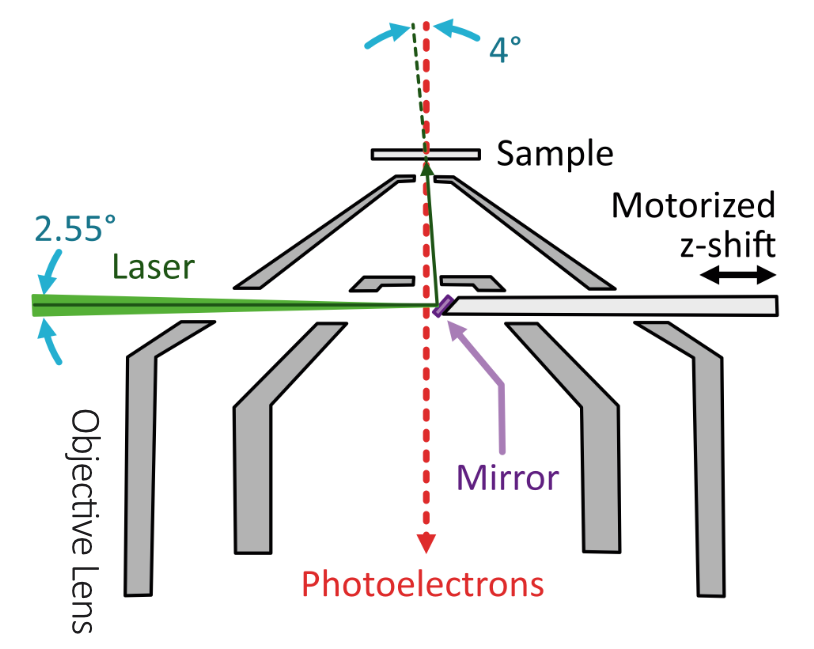
Normal Incidence Mirror
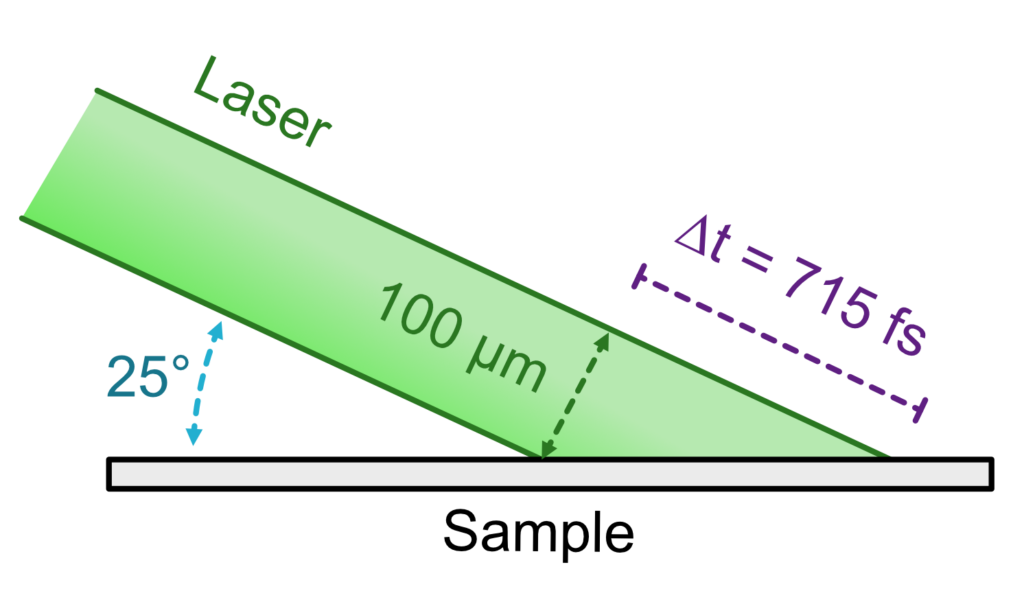
Under typical angles of incidence for a PEEM (25°) a laser with 100 µm spot size has a time difference of up to 715 fs when reaching the surface. This can be an issue for studying effects on the fs timescale.
A light-reflecting mirror can be integrated into the objective lens, which allows to excite the sample under nearly normal incidence. This way, the light pulse reaches the sample surface homogeneously at the same time.
A Modular Concept
TOF MARIS (time-of-flight energy filter) and NanoESCA MARIS (double-hemispherical energy filter) follow a modular concept, in which the microscopy lens can be ordered or upgraded with different energy filters. A popular set-up is to combine the MARIS front lens with a double-hemispherical energy-filter (suitable for a wide range of light sources) with a time-of-flight energy-filter (often an efficient choice for pulsed sources).
In accordance with the main scientific application, it is possible to start with a TOF MARIS and upgrade the double-hemispherical energy-filter at a later time, when additional experiments with continuous lightsources and a high flux become of interest. Vice versa, a NanoESCA MARIS can be upgraded with a time-of-flight energy filter. The double-hemispherical energy-filter can be upgraded with an Imaging Spin-Filter.
| Property | Specification |
|---|---|
| Momentum Space Resolution | < 0.005 Å-1 |
| Momentum Space Field of View | 0.5 Å-1 .. 6.0 Å-1 |
| Angular Acceptance Equivalent | ± 90° (full half sphere) |
| Real Space Resolution | 35 nm |
| Real Space Field of View | 5 µm .. 800 µm |
| Imaging Energy Resolution | 30 meV |
| Drift energy | 10 eV .. 100 eV |
| DLD Detector Time Resolution | 160 ps (< 100 ps achieved) |
| DLD Detector Count Rate (max.) | 1 MCounts/s |
| DLD Detector retractable | Yes, to use Standard Imaging Unit instead of DLD |
| Standard Imaging Unit | MCP/Camera for measurements with high dynamics (> 30 Mcount/s equivalent) |
| Contrast Aperture in Back Focal Plane | Discrete micro apertures, 5 fixed sizes |
| Grid in Back Focal Plane | Included, for Image Optimization |
| Field Aperture in first Image Plane | Continuously adjustable Iris aperture |
| High-Pass Filter for Std. Imaging Unit | Available as option (Integrated Energy Filter) |
| High Pass Filter for DLD | Available as option (Suppression of Secondary Electrons) |
| Normal Incidence Mirror | Available as option |
| Optional Field Aperture in first Image Plane | Discrete micro apertures, 5 fixed sizes |
| Grid in first Image Plane | Included in Optional Field Aperture, for Image Optimization |