SEM imaging with spin contrast
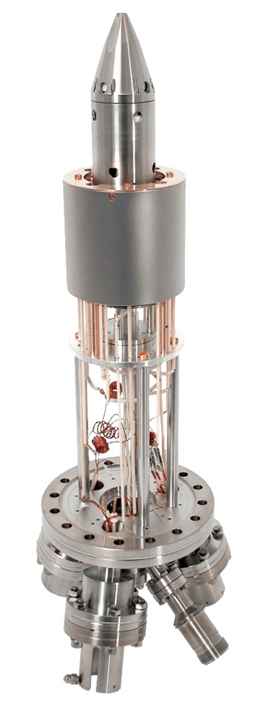
Scanning Electron Microscopy with Polarization Analysis (SEMPA) is a technique for directly imaging the magnetic structure of surfaces and thin films at the mesoscale by means of a scanning electron microscope, equipped with a suitable spin detector. A lateral resolution down to 10 nm is achievable in the SEMPA image with state of the art UHV SEMs. The FOCUS SEMPA detector is a dedicated spin detector that allows for parallel detection of two orthogonal spin channels. It is based on the well proven FOCUS SPLEED design. Combined with the ease of use of the SEM the FOCUS SEMPA detector provides a powerful tool to investigate electron transport in dependence of the pattern shape and magnetic domain structure. Alternatively the SEMPA detector has already been succesfully equipped with the FERRUM spin detector.
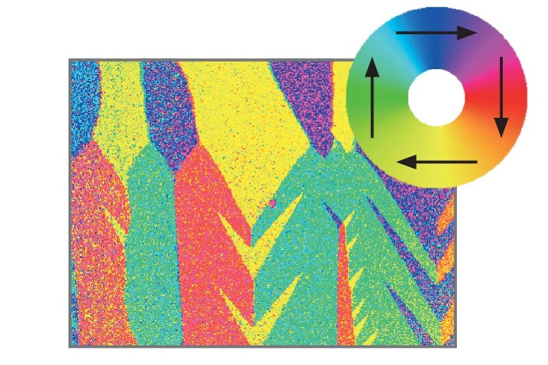
SEMPA image using Focus SPLEED of the magnetic domains of an iron whisker with a typical „fir tree“ structure. Each colour indicates a different direction of the magnetisation (4 different domain orientations) as indicated with the insert above.
Today the FERRUM spin detector is also available for use as a SEMPA detector.
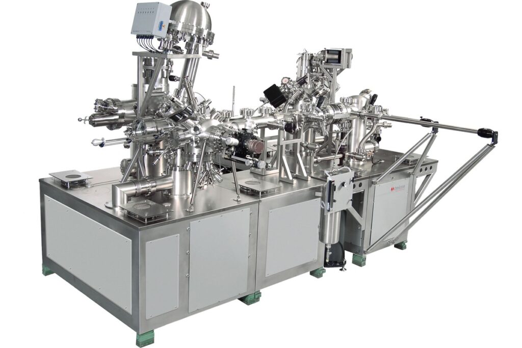
UHV Spintronics Cluster Tool (Scienta Omicron) that features a UHV Gemini based SEMPA system (SEM with polarization analysis) for magnetic imaging combined with FIB nano-structuring facilities.