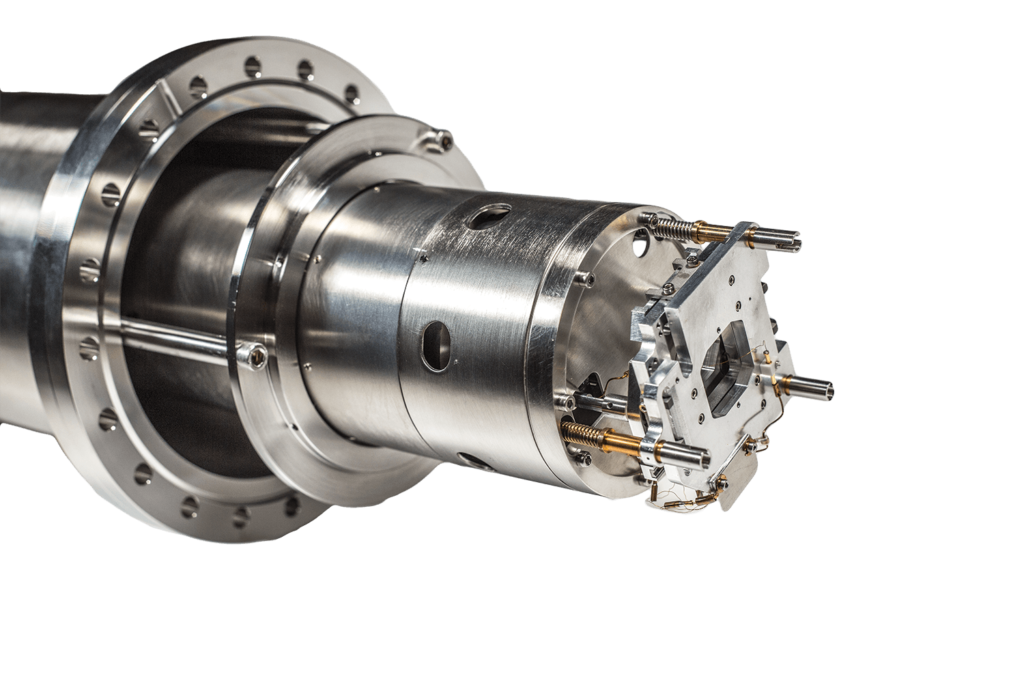
FOCUS IS-PEEM
Photoemission Electron Microscope with integral sample stage (IS) for unsurpassed stability and precise sample positioning via remote controlled piezo drives. The in-situ variable contrast aperture and the stigmator/deflector allow the power of PEEM to be fully exploited in laboratory and synchrotron applications. Topography contrast, work function contrast, chemical contrast and magnetic contrast can be used for surface sensitive real-time imaging of any reasonably flat and conducting surface.
The PEEM essentially consists of an imaging electrostatic lens system and a UV light source for the creation of photoelectrons via photoemission. The photoelectrons emitted from the surface are imaged onto a channelplate for amplification and finally onto a fluorescent screen. The image is acquired using a CCD camera typically. In contrast to a SEM, the PEEM does not use a scanned probe beam, but the sample surface is uniformly illuminated by, e.g., UV light or X-rays. This way beam induced damage of delicate surfaces is avoided. The magnified image of the surface can be observed directly and in real-time (even at video frequency if the photon intensity is sufficient) on the fluorescent screen.
In terms of it’s parallel image acquisition, the basic principle of operation is similar to an optical microscope. However, since electrons are used for imaging, the resolution is no longer limited by the wavelength of the photon beam. Instead, a strong electrostatic field between the sample and the objective lens accelerates the electrons released to energies of typically 10 to 15 keV. Thus a lateral resolution of up to 20 nm can be achieved.
PEEM
Photoemission Electron Microscope with integral sample stage (IS) for unsurpassed stability and precise sample positioning via remote controlled piezo drives. The in-situ variable contrast aperture and the stigmator/deflector allow the power of PEEM to be fully exploited in laboratory and synchrotron applications.
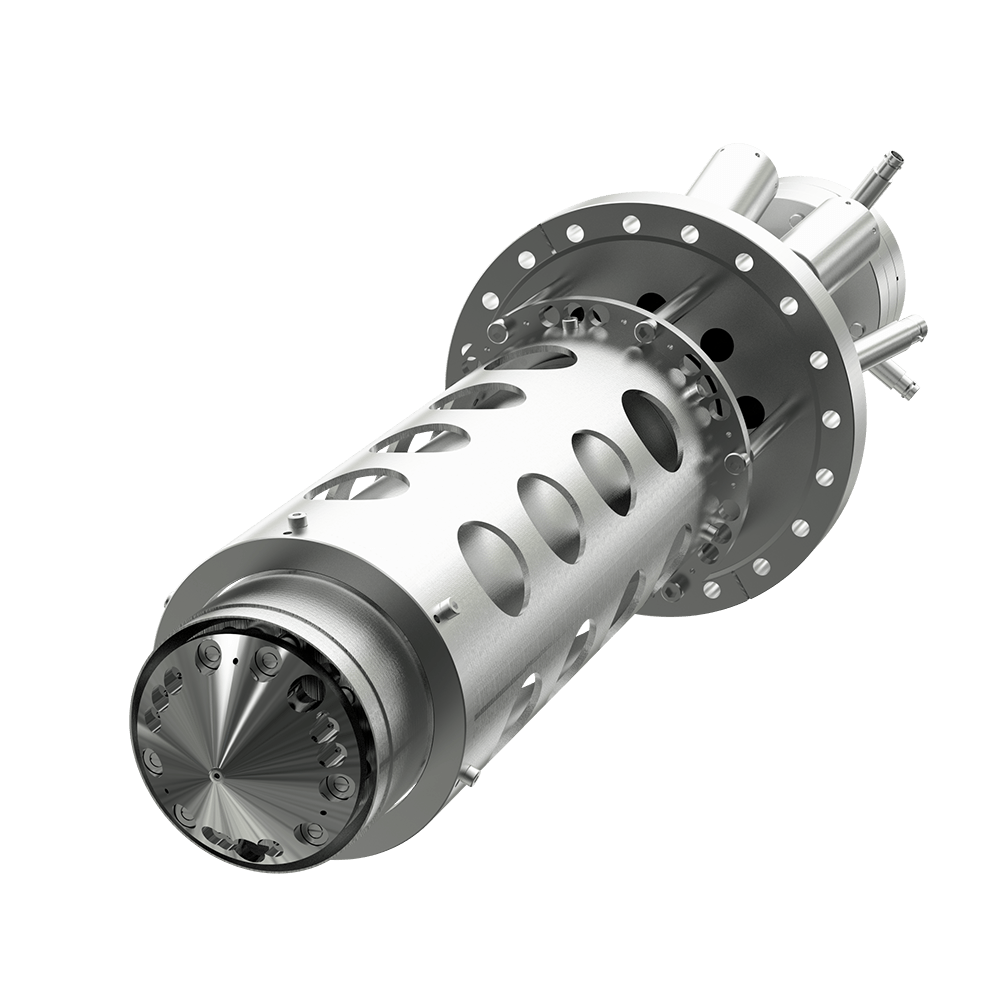
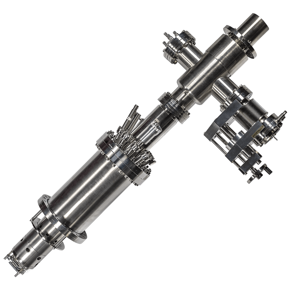
IEF / TOF PEEM
The Imaging Energy Filter (IEF) makes the FOCUS PEEM a powerful tool to measure work function maps (with lab sources) as well as element specific maps (with synchrotron, X-ray microspectroscopy) of conductive samples. For time/energy resolved measurements paired with a pulsed excitation source the FOCUS PEEM can be equipped with a Time-of-Flight (TOF) energy filter. All these options can be combined in one instrument for maximum versatility. The PEEM with IEF and/or TOF combined with the optional angular imaging optics delivers laterally resolved k-space images (momentum microscopy).
The FOCUS IS-PEEM is distributed by our sales partner Scienta Omicron. Please contact them for more informations.