NanoESCA with additional k-space lens:
The “Momentum Microscope”
The NanoESCA can be equipped optionally with additional electron optical elements to accomplish not only the lateral imaging but also the imaging of the angular distribution of the emitted electrons. This concept has been called Momentum Microscope.
360°-3d view of the Momentum Microscope
(NanoESCA with k-space optics):
Basically the NanoESCA consists of a complete PEEM optics completed by an imaging energy filter (IDEA) placed in between two projection lens sections. Finally the electrons are visualized typically by a multi channel plate/fluorescent screen unit together with a slow scan CCD camera.
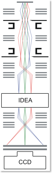
The electron optical approach is schematically shown here: Both figures show the electron optical ray tracing from the sample (top) to the imaging detector (bottom).
—————————————–>
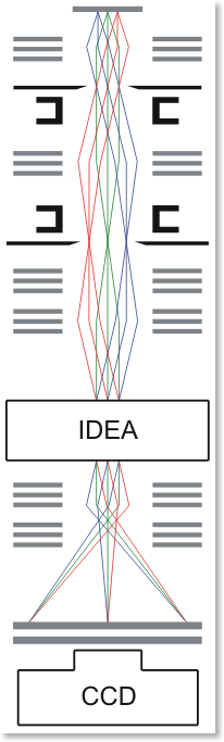
Real space imaging————————————>k-space imaging
The left hand figure illustrates the set-up causing a microscopic image of the real space (real space image). All electrons origin from real sample sites within the microscopic field of view (FOV) are energy filtered by the IDEA and imaged (magnified) on to the detector.
The right hand scheme represents the imaging of the angular distribution (k-space, momentum distribution) of the photo electrons emitted by the sample (k-space imaging).
By means of this unique electron optics it is possible to switch from real space imaging to k-space imaging and vice versa all the time. The angular distribution of the emitted electrons originating from a certain real space field of view are imaged for the kx and ky direction and a certain energy E at once. This kind of Micro-ARPES is illustrated nicely on a silver poly crystal as an example.
By sweeping the energy E of the imaged electrons it becomes possible to get the whole kx-ky-E image stack. This image stack represents a complete 3D-image of the band structure of the sample. The maximum FOV in the k-space depends upon the excitation energy applied. For HeI excitation (21.2eV) we achieve about 3 Å-1. Using HeII photons (40.8eV) this k-space FOV is expanded to about 6 Å-1.