FOCUS engagement with the PEEM dates back into 1996. Today almost 90 of our instruments are operated world wide. From the very first day we took all the customer feed-back to constantly improve the performance and ease of use of our microscopes. The FOCUS PEEM utilizes the technique of photoemission electron microscopy to image electrons emitted from any flat and conducting sample surface. Electron emission from surfaces can be caused by UV, VUV or x-ray photons, by thermal activation, by electron/ion bombardment or even by field emission. The FOCUS PEEM together with the dedicated high stability integrated sample stage (IS) and various available energy filters provides an easy to upgrade modular concept. Today the FOCUS PEEM is designed to combine both real- and k-space imaging together with energy filtering and hence spectroscopic microscopy („spectromicroscopy“) at its best.
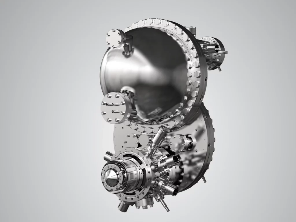
The most prominent energy filter is the patented IDEA. In combination with the PEEM known as NanoESCA .
Both the FOCUS IS-PEEM and the NanoESCA – also referred to as Momentum Microscope – you can purchase via Scienta Omicron as a component or turn-key system solution.
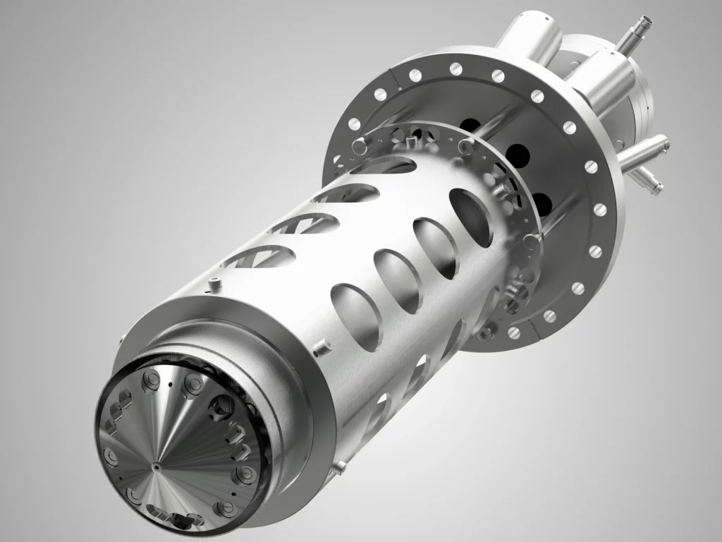
Photoemission Electron Microscope with integral sample stage (IS) for unsurpassed stability and precise sample positioning via remote controlled piezo drives. The in-situ variable contrast aperture and the stigmator/deflector allow the power of PEEM to be fully exploited in laboratory and synchrotron applications.
The Imaging Energy Filter (IEF) makes the FOCUS PEEM a powerful tool to measure work function maps (with lab sources) as well as element specific maps (with synchrotron, X-ray microspectroscopy) of conductive samples. For time/energy resolved measurements paired with a pulsed excitation source the FOCUS PEEM can be equipped with a Time-of-Flight (TOF) energy filter. All these options can be combined in one instrument for maximum versatility. The PEEM with IEF and/or TOF combined with the optional angular imaging optics delivers laterally resolved k-space images (momentum microscopy).
Next Generation Photoemission Tool for Real- and Momentum Microscopy.
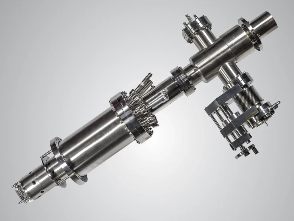
The core element consists of the patented IDEA (Imaging Dispersive Energy Analyser). Its double hemisphere design exhibits a real energy dispersive approach together with an unrevealed real and momentum space imaging quality (D. Funnemann, M. Escher, European Patent EP 1 559 126 B1, US patent US 7 250 599 B2).
The aberration compensated band pass filter IDEA does not only eliminate the analyser’s spherical aberration but the tandem arrangement also largely retains the time structure of the electron signal, unlike a single hemispherical analyser which can be helpful with time resolving experiments.
Click here for a 3d animation showcasing the working principle.
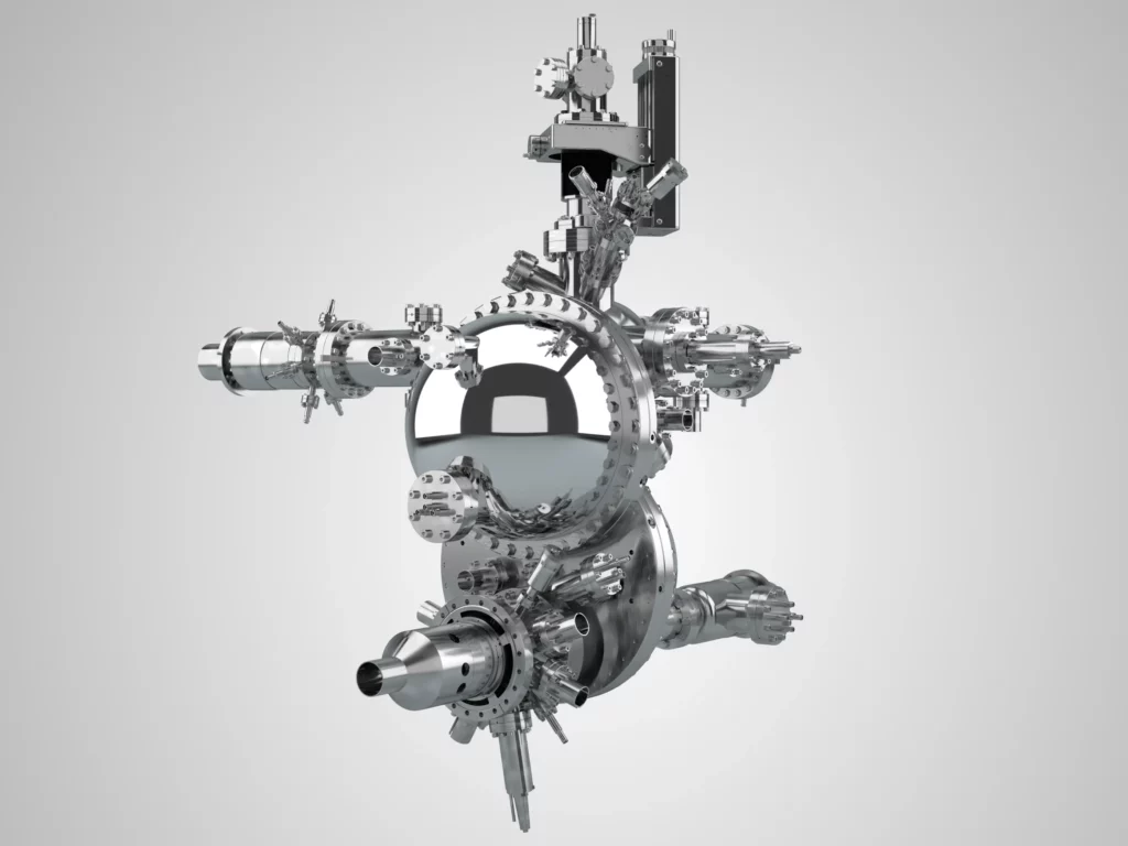
The NanoESCA can easily be equipped with the 2D Imaging Spinfilter. The NanoESCA III directly provides the real – and momentum space image at the required scattering energy of the 2D Imaging Spinfilter. This is a pre-requisite for un-compromised parallel acquisition of high resolution spin filtered images reducing measurement time by a factor of up to 10.000 compared to classical single channel detection.